Mapping the brain’s highways
New map showing messaging routes in the brain may help explain why some injuries are worse than others
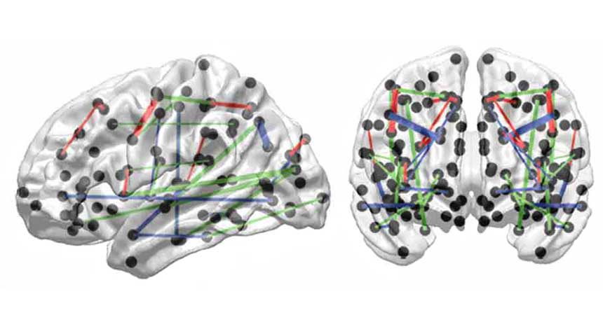
The green, blue and red lines show white matter pathways that connect different parts of the brain. This map was produced by scientists who used two different types of technologies to scan the brains of more than 100 healthy volunteers.
J. VAN HORN/FRONTIERS IN HUMAN NEUROSCIENCE 2014
Some people use gray matter as a slang term to mean intelligence or brains. Gray matter is in fact real, one of the two major types of tissues making up the brain. White matter is the other type. Like superhighways, its big bundles of nerve fibers connect different regions of gray matter. And like traffic, important messages travel along the white matter from one part of the brain to another. A new map of these superhighways suggests why some brain injuries are more devastating than others: It’s because they create a traffic jam — or outright roadblock — in a particularly important thruway.
Scientists published this new white-matter map on February 11 in Frontiers in Human Neuroscience.
In 1848, a man named Phineas Gage was working on a railroad in Vermont. Suddenly, an explosion drove a large iron rod straight through the front of his brain. It didn’t kill him (although his friends reported that afterward his personality was never the same).
The new map helps explain this. How much an injury impacts the brain seems to depend largely on how badly it damages the communications highways. “If a similar-sized injury to Mr. Gage had occurred in another location in his brain,” John Van Horn told Science News, “the damage to his network would have been much more severe.”
Van Horn is a neuroscientist at the University of Southern California, in Los Angeles. To create the new map, he and his coworker Andrei Irimia studied the brains of 110 healthy men. The scientists used two kinds of scans. One type, called MRI (for magnetic resonance imaging), revealed the brain’s basic anatomy. The other, called diffusion tensor imaging, showed the long tracts, or pathways, of nerve fibers that make up white matter. (These bundled fibers are sheathed in a protective layer of white insulation, which gives rise to the tissue’s name.)
The two experts used a computer to combine the information from both types of brain scans. The resulting map showed how white matter links different parts of the brain — almost like the wires in an electrical circuit.
Next, the scientists did some virtual snipping. They started severing connections to see which would hurt the flow of information most. Van Horn likened the process to cutting the wires, one by one, that carry electricity throughout a house. “Sometimes you’d cut a wire that doesn’t really change anything other than the outlet in your office,” he told Science News. “Other times you go and cut another wire, and it shuts off the power to your whole house.”
This approach revealed which connections, when broken, most harmed the relay of information. Some highways appear so important that even a minor injury would cause major problems. Other links may be less important, explaining why a seemingly major injury — like Mr. Gage’s — was not catastrophic.
The new map is not the first research to look at the connections in the brain. But most other ones had focused on gray matter, not the white-matter pathways.
The two approaches are “different ways of getting at the same problem,” Olaf Sporns told Science News. Sporns, a neuroscientist at Indiana University in Bloomington, did not work on the new map. However, he calls the new map “a very important extension” of previous research, including his own, on the connections within the brain.
Power Words
diffusion tensor imaging A technique for identifying microscopic changes or differences in the structure of brain regions. It can map the three-dimensional diffusion of water into tissues, which can change with disease. It’s often used to study the brain’s white matter.
gray matter One of two main types of tissue found in the brain and spinal cord. It consists mainly of nerve cell bodies.
magnetic resonance imaging (MRI) An imaging technique to visualize soft, internal organs, like the brain, muscles, heart and cancerous tumors. MRI uses strong magnetic fields to record the activity of individual atoms.
neuroscience Science that deals with the structure or function of the brain and other parts of the nervous system. Researchers in this field are known as neuroscientists.
tissue Any of the distinct types of material, comprised of cells, which make up animals, plants or fungi.
white matter One of the two main tissue types found in the brain and spinal cord. It consists mainly of bundles of nerve fiber.