Explainer: How to read brain activity
Scientists use a variety of tools to eavesdrop on electrical signaling in the brain
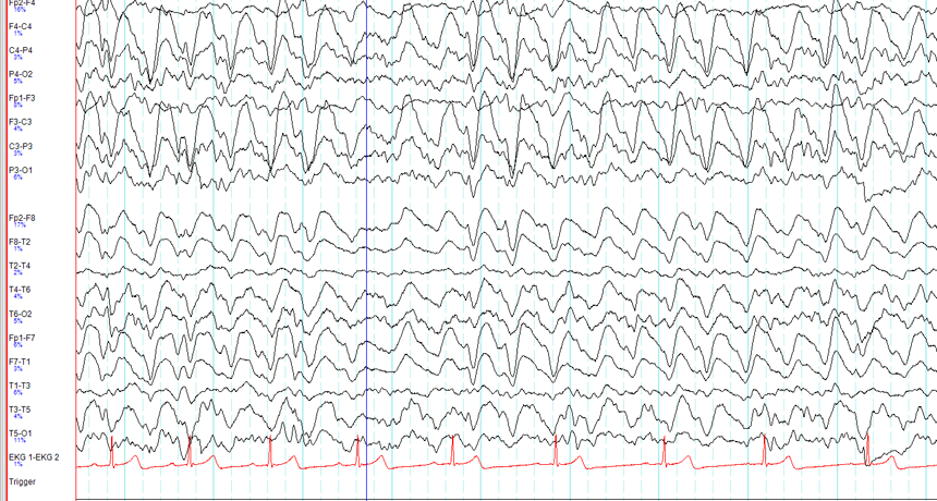
This graph of electrical activity in the brain is known as an EEG. Here, the patterns of spikes show that the person was asleep and likely dreaming.
Public domain
Neuroscientists study the inner workings of the human brain. To do this, they may listen in as tiny nerve cells, or neurons, communicate with each other. Their chatter consists of tiny zaps of electricity. These electrical signals form thoughts and feelings. They also control the body.
A single neuron fires off a jolt of electricity in response to a very specific trigger. Scientists call each jolt an action potential.
For instance, some nerve cells involved in vision fire only when the human eye sees certain colors. Others fire when the eye detects basic shapes, or the edges of objects.
Many action potentials combine to form bigger patterns of brain activity. These patterns become thoughts, movements and more. Imagine that those vision neurons detect a mix of colors and edges that look like a baseball, for example. Other networks of neurons may tell the arm to reach out and pick it up. Still other networks may form thoughts about playing catch.
These patterns of thought and movement, combine to form brainwaves. These are overall patterns of the electrical activity happening in the brain at any one time.
Observing the brain’s electrical activity can help scientists better understand how the brain works. Scientists use a variety of tools to measure action potentials, activity patterns and brainwaves.
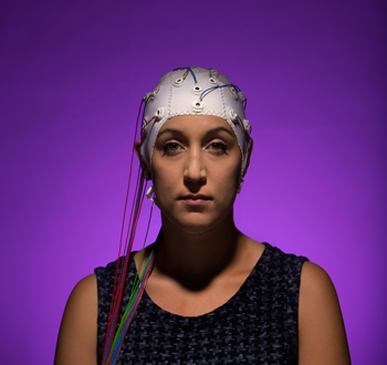
Helmets and headbands
The simplest way to study brain activity is to cover the head with a helmet, cap or headband dotted with flat metal sensors. These sensors can pick up electricity inside the brain. A device that graphs that activity is called an EEG, short for electroencephalograph (Ee-LEK-tro-en-SEF-uh-lo-graf).
An EEG can reveal if someone is awake, asleep or even dreaming. Awake, the brainwaves will look like a bunch of sharp spikes. As someone drifts off to sleep, those spikes will smooth into wavy hills.
EEG headsets also can give someone basic control over a computer program. To do this, a computer program needs to recognize the overall brainwave pattern of a few specific thoughts. Each thought might correspond to an action, such as pushing, tapping or swiping. Every time the person imagines one of those actions, the headset will read that thought as a command.
One such headset lets people use their thoughts to play simple computer games, control a toy car or browse through photos. Tan Le, who heads a company called Emotiv, demonstrated the technology in 2010. She gave a volunteer named Evan one of the headsets to try out. It recorded Evan’s brain activity as he looked at a floating cube on a computer screen. Evan imagined pulling on the cube. At first nothing happened. The computer program had to learn Evan’s brainwave patterns for pulling. Later, Evan imagined pulling again. This time, the cube sprang forward.
Electrodes in the brain
Unfortunately, EEG headsets can’t provide very detailed information. For a neuroscientist, using one to detect brain activity is like trying to hear an orchestra play from outside of the concert hall. You have to get closer to the musicians to figure out how the music is put together. In the brain, the closer a detector is to the individual neurons, the better it translates their electrical signals.
One type of detector has sensors that sit right next to the neurons themselves. To get them there, a surgeon must implant the tiny metal sensors inside the brain.
The brain has billions of neurons. It would be too dangerous to implant enough sensors to record all of them. But recording just a handful in one part of the brain can provide useful data. Imagine dropping a tiny microphone into the concert hall to record the notes played by just a few instruments. That might be enough to identify a song.
For example, in 2012, surgeons implanted two tiny detectors in the area of the brain that controls movement. Using the data they collected, Andrew Schwartz and a team of researchers at the University of Pittsburgh were able to translate patterns of action potentials into specific motions. Then, they set up a computer program to watch for these patterns. The program was connected to a robotic arm that would move according to the detected brain activity. This let a monkey or a human control the robotic arm using thought alone.
Sensors all over the brain
Electrodes inside the brain give an incredibly detailed view of electrical activity. But few people would volunteer to have their brains opened up and sensors put in.
Some people with rare brain diseases, however, already have a grid of electrical sensors implanted below their skulls, on their brains’ surface. Those sensors read brain activity through a technology called electrocorticography (Ee-LEK-tro-KOR-tih-KOG-rah-fee), or ECoG.
Each ECoG sensor eavesdrops on hundreds of thousands of neurons at once. This is like listening to the concert from the back of the auditorium. It’s easy to hear the tune. But it’s harder to tease apart which notes are coming from which instruments.
Magnets for brain mapping
Scientists have yet another way to spy on brain activity. It requires no surgery, yet provides more detailed data than do EEGs. Unlike the other three methods, this one does not directly record electrical activity. Instead, it tracks blood flow in the brain.
Imagine not being able to hear the concert at all. You can only watch a silent video of the musicians. Even without sound, seeing the musicians’ arms and fingers move would give you clues about the music they’re playing. In a similar way, functional magnetic resonance imaging — or fMRI — maps which brain cells are especially active.
Volunteers lie down on a table that slides inside a large, donut-shaped machine. There, magnets measure the blood flow that carries chemical food to neurons. Actively firing neurons suck up the most food. So mapping where blood flow is highest points to where the brain is most active.
Each of these technologies has its own benefits and drawbacks. Scientists choose between them based on the type of information they seek. They may get new options as engineers continue to improve the sensors themselves. Eventually, devices will likely become advanced enough to translate patterns of brain activity into words, pictures and more.